
|
|
[China]

Trade Verify
Address: Room 1705,Office Building B1,Wanda Plaza,No.2707,Kaichuang Road,Guangzhou,Guangdong,China
Contact name:MileyZhu

Guangzhou Rongtao Medical Tech LTD. |
|
Verified Suppliers
|
|
|
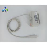
Hitachi EUP-S72 Phased Array Ultrasound Transducer Scanner Probe For Cardiology
1. Type: Phased Array
2. Applications: Cardiology
3. Frequency Range: 2 - 9 Mhz
4. Compatibility: HI VISION, EUB
Other Hitachi Aloka Ultrasound Probes we can offer:
| Brand | Model |
| Hitachi Aloka | EUP-B512 |
| Hitachi Aloka | BUP-B712 |
| Hitachi Aloka | EUP-S70 |
| Hitachi Aloka | EUP-S72 |
| Hitachi Aloka | EUP-V73W |
| Hitachi Aloka | L441 |
| Hitachi Aloka | S211 |
| Hitachi Aloka | UST-52126 |
| Hitachi Aloka | UST-5299 |
| Hitachi Aloka | UST-5412 |
| Hitachi Aloka | UST-5413 |
| Hitachi Aloka | UST-9118 |
| Hitachi Aloka | UST-9124 |
| Hitachi Aloka | UST-984-5 |
| Hitachi Aloka | EUP-L74M |

Knowledge Point
Multi-frequency probe
Multi-frequency probe is a new development of pulse-echo transducer. The probe can send out several different ultrasonic pulses, so as to cover the near field with high-frequency ultrasound, the intermediate-frequency ultrasound to cover the transition zone between the far and near fields, and the low-frequency ultrasound to cover the far field. The unit multi-frequency probe is to bond multiple layers of piezoelectric ceramics (or polymer piezoelectric materials) to each other, and lead wires from the electrodes between each layer to obtain multiple frequency ultrasonic pulse emission through exciting different layers . The digital coding of the multi-frequency probe is simple but easy to lose the signal.
1. Sonogram pictures can help identify the basic anatomy of a developing fetus, and can often detect birth defects and abnormalities very early on.
2. Because of these test results, there is ongoing research into the possible ill effects of sonograms on human fetuses. While many people regard sonograms as a test to determine the gender of an unborn child, the procedure is more often utilized to monitor the progress of development, or to identify the origin of some unusual pain or discomfort experienced by the mother, such as pain accompanied by bleeding.
Storage for Transport of ultrasonic transducer
1. Make sure that the transducer is clean and disinfected before placing it in the case to avoid contaminating the foam that lines the carrying case.
2. Place the transducer in the case carefully to prevent kinking of the cable.
3. Before closing the lid, make sure no part of the transducer is protruding from the case.
4. Wrap the case in plastic material containing sealed-air pockets (such as Bubble Wrap material), and pack the wrapped case in a cardboard carton.


