

Active Member
|
[China]

Address: RM718, EASTERN BUILDING, XIAOBEI, YUEXIU, GUANGZHOU, CHINA.
Contact name:Andy

Guangzhou MeCan Medical Limited |
|
|
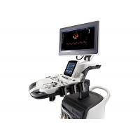
The Most High-End Trolley Color Doppler ECHO Ultrasound Scanner For Hospital
Moidel: S40
Feasures of the Ecocardiografos Doppler Color :
Through years of continuous innovation, development, and by giving
priority to our customers’ requirements, is proud to release its
new generation, high-level ultrasound product, S40. Based on a
revolutionary new platform, combining SonoScape's core imaging
technologies and sleek ergonomic design, the S40 represents the new
standard of SonoScape S series products. This elevates imaging
performance to a record level and satisfies even the most demanding
clinical requirements, significantly expanding the value of
ultrasound.
19 inch widescreen
10 inch smart interactive graphical Touch Screen
High Resolution Monitor
-- High definition 1024*768
--Wide viewing angle for 170°
Four-way Articulated Arm with lock, vertical and horizontal
rotation
Fully adjustable and rotating Control Panel
User-oriented multinational language input keyboard
Gel Warmer and Endocavity Probe Holder
5 Transducer Sockets plus one special connector for Pencil Probe
Full range of Transducers configured with latest technologies
Patient-oriented File Management System.
Flexible user-defined functions
Customized one button functions
Optional Wi-Fi and Blue Tooth functions
Full patient database solution:DICOM3.0, AVI/JPG, USB2.0,
HDD,DVD,PDF report
Premium Platform with Perfect Work Flow
S40 is committed to providing doctors with exceptional imaging
quality in both near and far fields, greatly improving resolution
and penetration for greater diagnostic confidence.
u-Scan
u-Scan Technology uses real-time image processing algorithms to
eliminate speckle and noise artifacts, enhancing tissue margins and
borders by correcting discontinuity between different regions. Thus
allowing improved visualization of real tissue information. It can
also automatically adapt the acoustic velocity in different regions
to improve the resolution and contrast.
Spatial Compound Imaging
Spatial Compound Imaging utilizes several lines of sight for
optimal contrast resolution, speckle reduction and border
detection. Configured with SCI technology, the S40 is ideal for
superficial and abdominal imaging with better clarity and improved
continuity of structures.
Pulse Inversion Harmonic Imaging
| 128 elements linear array L741(Vascular, Small parts, MSK etc.), 4-16MHz/46mm |
| 192 elements linear array L742(Vascular, Small parts, MSK etc.), 4-16MHz/ 38mm |
| 256 elements linear array L752(Vascular, Small parts, MSK etc.), 4-16MHz/ 52mm |
| 128 elements convex array C344 (Abdominal, Obstetrics, Gynecology), 2-6.8MHz/ R40mm |
| 128 elements convex array C322 (Abdominal, Biopsy), 2-6.8MHz/ R20mm |
| 192 elements convex array C353 (Abdominal, Obstetrics, Gynecology), 2-6.8MHz/ R50mm |
| 128 elements convex array C611 (Pediatric), 4-13MHz/ R8mm |
| 80 elements phased array 2P2 (Cardiac, Transcranial), 1-6MHz |
| 80 elements phased array 3P1 (Cardiac, Transcranial), 1-6MHz |
| 96 elements phased array 5P2 (Cardiac, Transcranial, Pediatric), 3-9MHz |
| 96 elements phased array 8P1 (Cardiac, Transcranial, Pediatric), 4-12MHz |
| 128 elements endocavity 6V1 (Gynecology, Obstetrics, Urology), 3-15MHz/ R11mm |
| 192 elements endocavity 6V3 (Gynecology, Obstetrics, Urology), 3-15MHz/ R10mm |
| 128 elements endocavity EC9-5 (Urology, Gynecology, Obstetrics), 3-15MHz/ R8mm |
| Volumetric convex array VC6-2 (Obstetrics, Abdominal, Gynecology), 2.2-6MHz/ R40mm |


