
|
|
[China]

Trade Verify
Address: Room 1705,Office Building B1,Wanda Plaza,No.2707,Kaichuang Road,Guangzhou,Guangdong,China
Contact name:MileyZhu

Guangzhou Rongtao Medical Tech LTD. |
|
Verified Suppliers
|

|
|
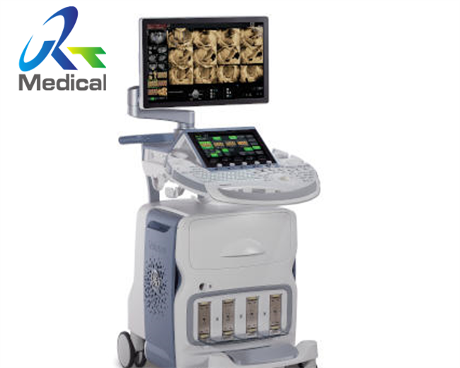
GE Voluson E8 BT15 SATA Data Cable for HD Drive KTZ300244 Medical Equipment Supply
1. Part number: KTZ300244
2. Compatible with Voluson E8
3. New original
4. Warranty: 90 days
5. Lead time: 2-4 days
Various parts of GE Voluson E8 we can offer:
| System | Part Number | Description |
| Voluson E8 BT15 | KTZ304229 | RTU100 Console |
| Voluson E8 BT15 | KTZ303892 | RSP3-3C-Power Supply EC300 |
| Voluson E8 BT15 | KTZ3a03954 | RTF200-EC300 Probe Control Board |
| Voluson E8 BT15 | KTZ303915 | RFM221 |
| Voluson E8 BT15 | KTZ303916 | RFM201 |
| Voluson E8 BT15 | KTZ303054 | RSX20 |
| Voluson E8 BT15 | KTZ303250 | RSX10 |
| Voluson E8 BT15 | KTZ304029 | Graphic Card 5 EC300 |
| Voluson E8 BT15 | KTZ303830 | Back End Processor(BEP)Kit BT15 |
| Voluson E8 BT15 | KTZ280290 | RTT3.P3-Distribution Board TOP |
| Voluson E8 BT15 | KTZ304040 | RTH50-Distribution Board |
| Voluson E8 BT15 | KTZ303930 | RTB50.P1 Distribution Board BOTTOM |
| Voluson E8 BT15 | KTZ303517 | RTV30 |
| Voluson E8 BT15 | KTZ300244 | SATA Data Cable for HD Drive |
| Voluson E8 BT15 | KTZ303121 | PCI-E Connection Cable |
Knowledge point
Ultrasound apply in:
In anesthesiology, ultrasound is commonly used to guide the placement of needles when placing local anaesthetic solutions near nerves. It is also used for vascular access such as central venous cannulation and difficult arterial cannulation. Transcranial Doppler is frequently used by neuro-anesthesiologists for obtaining information about flow-velocity in the basal cerebral vessels.
Further information: Doppler ultrasonography and Intravascular ultrasound
In angiology or vascular medicine, duplex ultrasound (B Mode imaging combined with Doppler flow measurement) is used to diagnose arterial and venous disease. This is particularly important in neurology, where carotid ultrasound is used for assessing blood flow and stenoses in the carotid arteries, and transcranial Doppler is used for imaging flow in the intracerebral arteries.
Intravascular ultrasound (IVUS) uses a specially designed catheter with a miniaturized ultrasound probe attached to its distal end, which is then threaded inside a blood vessel. The proximal end of the catheter is attached to computerized ultrasound equipment and allows the application of ultrasound technology, such as piezoelectric transducer or CMUT, to visualize the endothelium (inner wall) of blood vessels in living individuals.
In the case of the common and potentially, serious problem of blood clots in the deep veins of the leg, ultrasound plays a key diagnostic role, while ultrasonography of chronic venous insufficiency of the legs focuses on more superficial veins to assist with planning of suitable interventions to relieve symptoms or improve cosmetics.
Echocardiography is an essential tool in cardiology, assisting in evaluation of heart valve function, such as stenosis or insufficiency, and strength of cardiac muscle contraction. such as hypertrophy or dilatation of the main chambers. (ventricle and atrium)
Point of care emergency ultrasound has many applications in emergency medicine. This includes differentiating cardiac causes of acute breathlessness from pulmonary causes, and the Focused Assessment with Sonography for Trauma (FAST) exam for assessing significant hemoperitoneum or pericardial tamponade after trauma. Other uses include assisting with differentiating causes of abdominal pain such as gallstones and kidney stones. Emergency Medicine Residency Programs have a substantial history of promoting the use of bedside ultrasound during physician training.
Abdominal and endoanal ultrasound are frequently used in gastroenterology and colorectal surgery. In abdominal sonography, the solid organs of the abdomen such as the pancreas, aorta, inferior vena cava, liver, gall bladder, bile ducts, kidneys, and spleen are imaged. However, sound waves are blocked by gas in the bowel and attenuated to differing degrees by fat, sometimes limiting diagnostic capabilities in this area. The appendix can sometimes be seen when inflamed (as in e.g.: appendicitis) and ultrasound is the initial imaging choice, avoiding unnecessary radiation, although it frequently needs to be followed by other imaging methods such as CT. Endoanal ultrasound is used particularly in the investigation of anorectal symptoms such as fecal incontinence or obstructed defecation. It images the immediate perianal anatomy and is able to detect occult defects such as tearing of the anal sphincter. Ultrasonography of liver tumors allows for both detection and characterization.
Gynecologic ultrasonography examines female pelvic organs (specifically the uterus, ovaries, and Fallopian tubes) as well as the bladder, adnexa, and Pouch of Douglas. It commonly uses transducers designed for approaches through the lower abdominal wall, curvilinear and sector, and specialty transducers such as endovaginal.
Obstetrical sonography was originally developed in the late 1950s and 1960s by Sir Ian Donald and is commonly used during pregnancy to check on the development and presentation of the fetus. It can be used to identify many conditions that could be potentially harmful to the mother and/or baby possibly remaining undiagnosed or with delayed diagnosis in the absence of sonography. It is currently believed that the risk of leaving these conditions undiagnosed is greater than the small risk, if any, associated with undergoing an ultrasound scan. But its use for non-medical purposes such as fetal "keepsake" videos and photos is discouraged.
Obstetric ultrasound is primarily used to:
According to the European Committee of Medical Ultrasound Safety (ECMUS)
Ultrasonic examinations should only be performed by competent personnel who are trained and updated in safety matters. Ultrasound produces heating, pressure changes and mechanical disturbances in tissue. Diagnostic levels of ultrasound can produce temperature rises that are hazardous to sensitive organs and the embryo/fetus. Biological effects of non-thermal origin have been reported in animals but, to date, no such effects have been demonstrated in humans, except when a micro-bubble contrast agent is present.
Nonetheless, care should be taken to use low power settings and avoid pulsed wave scanning of the fetal brain unless specifically indicated in high risk pregnancies.
Ultrasound scanners have different Doppler-techniques to visualize arteries and veins. The most common is color Doppler or power Doppler, but also other techniques like b-flow are used to show blood flow in an organ. By using pulsed wave Doppler or continuous wave Doppler blood flow velocities can be calculated.
Figures released for the period 2005–2006 by the UK Government (Department of Health) show that non-obstetric ultrasound examinations constituted more than 65% of the total number of ultrasound scans conducted.
Blood velocity can be measured in various blood vessels, such as middle cerebral artery or descending aorta, by relatively inexpensive and low risk ultrasound Doppler probes attached to portable monitors. These provides non-invasive or transcutaneous (non-piecing) minimal invasive blood flow assessment. Common examples are, Transcranial Doppler, Esophogeal Doppler and Suprasternal Doppler.
Most structures of the neck, including the thyroid and parathyroid glands, lymph nodes, and salivary glands, are well-visualized by high-frequency ultrasound with exceptional anatomic detail. Ultrasound is the preferred imaging modality for thyroid tumors and lesions, and ultrasonography is critical in the evaluation, preoperative planning, and postoperative surveillance of patients with thyroid cancer. Many other benign and malignant conditions in the head and neck can be evaluated and managed with the help of diagnostic ultrasound and ultrasound-guided procedures.
In neonatology, transcranial Doppler can be used for basic assessment of intracerebral structural abnormalities, bleeds, ventriculomegaly or hydrocephalus and anoxic insults (Periventricular leukomalacia). The ultrasound can be performed through the soft spots in the skull of a newborn infant (Fontanelle) until these completely close at about 1 year of age and form a virtually impenetrable acoustic barrier for the ultrasound. The most common site for cranial ultrasound is the anterior fontanelle. The smaller the fontanelle, the poorer the quality of the picture.
In ophthalmology and optometry, there are two major forms of eye exam using ultrasound:
Modern ultrasound is used to assess the lungs in a variety of settings including critical care, emergency medicine, trauma surgery, as well as internal medicine. This imaging modality is used at the bedside to evaluate a number of different lung abnormalities as well as to guide procedures such as thoracentesis, pleural drainage, needle aspiration biopsy, and catheter placement.
Pictures
FAQ
Q: Is there any guarantee for the goods?
A: Fully-tested and safe packing before shipping, less
than 3% warranty return claims of previous orders.
Q: Will you provide warranty?
A: Items are sold with NO WARRANTY, unless it is stated otherwise in the body of the proforma invoice/purchase order. Mostly 90 days for new original, 30/60 days for used.
Q: If faults occurred after warranty date, can I contact you?
A: Sure, feel free to contact even years later. We'll look into the
faults and try to find out some solutions/suggestions for you
reference.
Q: How about the payment terms?
A: All orders are 100% prepaid before delivery. We accept wire
transferring / Western Union from bank, and cash. (US dollars,
Euro, or CNY)
Q: How about delivery?
A: We ship worldwide. DHL/EMS/FedEx Express will be arranged within 1-5 working days after payment confirmed unless otherwise specified. We Seller are responsible for export Customs clearance in our end, and buyers for import Customs clearance in their end.


