

Active Member
|
[China]

Address: #1008.Junjiang Internatioanl Bldg. 228 Ningguo Rd. Yangpu Dist. Shanghai. 200090. China
Contact name:

Shanghai Korain Biotech Co., Ltd |
|
|
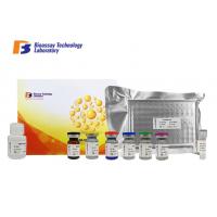
Porcine A1-Acid Glycoprotein Sandwich Test Kit High Specificity
A1-AGP ELISA Assay kit
Cat.No E0063Po
Reagent Provided
| Components | Quantity |
| Standard Solution (6400mg/L) | 0.5ml x1 |
| Pre-coated ELISA Plate | 12 * 8 well strips x1 |
| Standard Diluent | 3ml x1 |
| Streptavidin-HRP | 6ml x1 |
| Stop Solution | 6ml x1 |
| Substrate Solution A | 6ml x1 |
| Substrate Solution B | 6ml x1 |
| Wash Buffer Concentrate (30x) | 20ml x1 |
| Biotinylated Porcine A1-AGP Antibody | 1ml x1 |
| User Instruction | 1 |
| Plate Sealer | 2 pics |
| Zipper bag | 1 pic |
Standard Curve Range: 30mg/L - 6000mg/L
Sensitivity: 16.12mg/L
Size: 96 wells
*This product is for research use only, not for use in diagnosis
procedures. It’s highly recommend to read this instruction entirely
before use.
Precision
Intra-Assay Precision (Precision within an assay) Three samples of
known concentration were tested on one plate to assess intra-assay
precision.
Inter-Assay Precision (Precision between assays) Three samples of
known concentration were tested in separate assays to assess
inter-assay precision.
CV(%) = SD/mean x 100
Intra-Assay: CV<8%
Inter-Assay: CV<10%
Intended Use
This sandwich kit is for the accurate quantitative detection of
Porcine A1-Acid Glycoprotein (also known as A1-AGP) in serum,
plasma, cell culture supernates, cell lysates, tissue homogenates.
Precautions
Reagent Preparation
All reagents should be brought to room temperature before use.
Standard Reconstitute the 120μl of the standard (6400mg/L) with 120μl of
standard diluent to generate a 3200mg/L standard stock solution.
Allow the standard to sit for 15 mins with gentle agitation prior
to making dilutions. Prepare duplicate standard points by serially
diluting the standard stock solution (3200mg/L) 1:2 with standard
diluent to produce 1600mg/L, 800mg/L, 400mg/L and 200mg/L
solutions. Standard diluent serves as the zero standard(0 mg/L).
Any remaining solution should be frozen at -20°C and used within
one month. Dilution of standard solutions suggested are as follows:
| 3200mg/L | Standard No.5 | 120μl Original Standard + 120μl Standard Diluent |
| 1600mg/L | Standard No.4 | 120μl Standard No.5 + 120μl Standard Diluent |
| 800mg/L | Standard No.3 | 120μl Standard No.4 + 120μl Standard Diluent |
| 400mg/L | Standard No.2 | 120μl Standard No.3 + 120μl Standard Diluent |
| 200mg/L | Standard No.1 | 120μl Standard No.2 + 120μl Standard Diluent |
| Standard Concentration | Standard No.5 | Standard No.4 | Standard No.3 | Standard No.2 | Standard No.1 |
| 6400mg/L | 3200mg/L | 1600mg/L | 800mg/L | 400mg/L | 200mg/L |
Wash Buffer Dilute 20ml of Wash Buffer Concentrate 30x into deionized or
distilled water to yield 600 ml of 1x Wash Buffer. If crystals have
formed in the concentrate, mix gently until the crystals have
completely dissolved.
Summary
1. Prepare all reagents, samples and standards.
2. Add sample and ELISA reagent into each well. Incubate for 1 hour
at 37°C.
3. Wash the plate 5 times.
4. Add substrate solution A and B. Incubate for 10 minutes at 37°C.
5. Add stop solution and color develops.
6. Read the OD value within 10 minutes.
Calculation of Result
Construct a standard curve by plotting the average OD for each
standard on the vertical (Y) axis against the concentration on the
horizontal (X) axis and draw a best fit curve through the points on
the graph. These calculations can be best performed with
computer-based curve-fitting software and the best fit line can be
determined by regression analysis.


