|
|
[China]

Trade Verify
Address: Room 408, No.801 Hongqiao road, Wuxi, Jiangsu, China
Contact name:Helen Bo

Wuxi Biomedical Technology Co., Ltd. |
|
Verified Suppliers
|
|
|
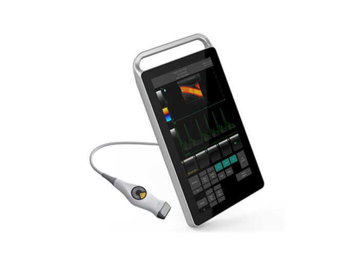
Trolley Color Ultrasound Scanner Color Doppler Machine With 18.5" Medical Touch Monitor
All touch-screen Color Doppler Ultrasound System
Advantage
1. 18.5-inch full touch screen design,to ensure aseptic operation.
2. All metal shell with unique design, sturdy and durable.
3. Professional software packages for Puncture application.
4. Build-in large capacity battery,for 8 hours continuous work.
5. Advanced image processing technology with high resolution display.
6. Multiple application:Reproductive Medicine,puncture guided,some special application like foot and Musculoskeletal system check, limited space clinic.
7. With special attention to mode,P6 include two different mode,Expert mode and Simple mode,suitable for any department and quite accessible for any doctor.
Function
-1 probe connector, which can be expand to 3 connectors by the probe expansion module
-18.5"high-resolution color LCD monitor
-Equipped with more than 4 beamformer function
-Equipped with THI (Tissue Harmonic Imaging), and second digital THI function
-Equipped with TSI (Tissue Specific Imaging) function
-Frequency compound imaging: adjustable under 2D and M modes
-Space compound imaging: adjustable under 2D and M modes with more than 3 levels
"-The scanning modes include 2D (2D ultrasound scanning diagnostic method), M (time motion, M mode diagnostic method), PW
(Pulsed Wave Doppler), CFM (Color Flow Mapping), PDI (Power Doppler Imaging)."
-Different frequencies are selected for 2D image and color image
-Equipped with biopsy guide line, guide by convex probe, transvaginal and linear probe
"-Equipped with bidirectional cine-loop function, gray scale cine-loop no less than 1024 frames, PW cine-loop time is no less than
100 seconds. Automatic/manual playback. The speed of the playback can be adjusted"
-Massive image storage (related with configured hard disk, no less than 500G)
-Equipped with 123 types of body marks; the probe position and scanning direction can be shown with arrow
Specification
| Monitor | 18.5" LCD touch monitor |
| Video Output | PAL-D, S-video, NTSC, VGA, DVI |
| Digital Scan Converter | 628× 440 × 24 bits |
| Body Mark | 123 body marks with probe location |
| Probe Frequency | 2. 0MHz~10. 0MHz |
| Gray Scale | 256 |
| Color Scale | 24 |
| Frame Rate | Max. up to 70 f/s |
| Scanning Area | ≥ 320 mm |
| Density of Scanning Lines | Max 256 line/frame |
| Cine loop | ≥1024 frames |
| Biopsy | Optional biopsy guide |
| Display Mode | B, 2B, 4B, M, B+M, CFM, PDI, B+PW, B+CFM+PW, B+PDI+PW |
| Type of Focusing | Dynamic focusing, acoustic lens focusing, launching multipoint focusing |
| Scanning Technique | Dynamic apodization, dynamic aperture, dynamic frequency scanning, multi-acoustic beam |
| Pre-processing | low noise preamplifier, TGC, filtering, frame average, line average |
| Post-processing | Gamma correction, histogram, digital scan conversion (DSC), edge enhancement, noise rejection, smooth, ePure, grey scale transformation, pseudo-color, color persistence etc |
| Display Control | Freeze/Unfreeze, Left-right reverse, Up-down reverse, Polarity reverse, picture rotation (90°/180°/270°), color reverse, inverse-frequency spectrum, pseudo-color. |
| Control of Transformation and Adjustment on Sound Field | output acoustic power, PRF, focus position, scanning angle, image frame frequency, impulse duration, depth, sampling area dimension |
| General Measurement | 2D/CFM: Distance, area, volume (ellipse method), angle; M: Distance, time, slope, heart rate, simplified left ventricle function, complete left ventricle function; PW: Distance, max ventricular gradient, average ventricular gradient, time, S/D ration, blood flow rate, acceleration of blood flow, heart rate, pulsate & drag index. |
| Image Memory | Hard disk for massive image storage, min. 10,000 images permanent stored |
| Character | Date, time, patient name, device name, user name etc, user-defined annotation table, arrowhead and body marks |
| Measurement Software | Software packages of Abdomen, Urology, Cardiology, Obstetrics, Gynecology, Superficial Parts, Blood Vessels, Fetal Heart, Transcranial Doppler and Orthopedic Surgery; directly form diagnostic report based on measurement results. |
| Input Power | 120VA |
| Continuous Working Time | >8 h |
| Size(3 cartons) | 65*42*18cm;140*15*9cm;73*60*39cm |
| Weight | 40kg |