|
|
[China]

Trade Verify
Address: 2nd floor of building #5, 27 Fengsheng Road, Jinfeng Town,High-Tech Industrial Development Zone, 401329 Chongqing, People's Republic of China
Contact name:Robert Wang

Chongqing Bio Newvision Medical Equipment Ltd. |
|
Verified Suppliers
|
|
|
retinal imaging camera capture the anterior and posterior eye images small pupil mode
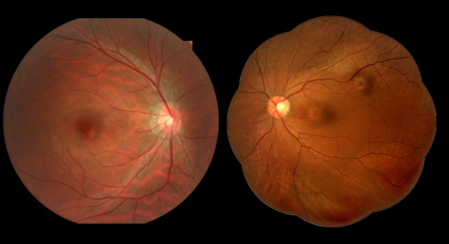
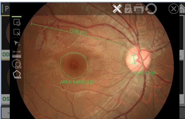
Digital Fundus Camera RetiCam 3100 is to take photos of the fundus of the eye through a fundus camera. Can clearly see the retinal blood vessels, macular area, optic nerve, is of great significance to the diagnosis of various types of eye disease, check is simpler, a few minutes to complete, no pain, there is a vision loss could be presbyopia may also be ophthalmology, need through the slit lamp examination and fundus examination can be confirmed.
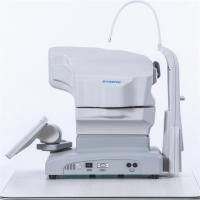
Digital Fundus Camera Reticam 3100 is a highly recognized and popular automatic fundus imaging system around the world. Through dual camera imaging, image control and feature recognition, eye XYZ 3D automatic positioning is realized. Through CCD imaging feedback, the exposure is measured, and the exposure intensity is automatically and accurately determined, without the need for a doctor's complex operation. By controlling the focusing step length according to the image resolution, the image definition can be automatically adjusted to obtain a clear and accurate fundus image and help doctors make accurate judgments for patients.Digital Fundus Camera RetiCam 3100 can be used to capture the anterior and posterior eye images. It has a field of view of 50 degrees for fundus imaging.
| Field of view | 50° |
| Field of view tolerance | ±7% |
| Resolution | |
| Center of view | ≥60 lp/mm |
| Field of view center (r/2) | ≥40 lp/mm |
| At the edge of the field of view (r) | ≥25 lp/mm |
| Magnification | 1.3 times |
| Required pupil diameter | 4.0 mm or more (3.3 mm or more when using the small pupil shooting function) |
| Working distance | 35 mm |
| Focus adjustment range | ±25D |
| Shooting light | Auto: with shooting mode |
| Manual: can be set manually | |
| light source | |
| Illumination light | Infrared led |
| Shooting light | Xenon lights |
| reflected light | |
| Scattered light | |
| camera | Digital camera |
| Fixing light | Internal fixing light (LED) |
| External fixation light | |
| Moving range | |
| Stage | 90 mm left and right, 35 mm front and rear |
| Main unit moves vertically | 30 mm |
| Chin-rest tray movement range | 60 mm |
| Rated power supply | AC 100V~240V,50/60Hz |
| size | 380 mm(L)x 550 mm(W)x 475 mm(H) |
| weight | About 26.5kg |
| When in maximum light intensity/hole bar state | |
| Spectral wavelength | 305 nm~1100 nm |