Home Companies Henan Aile Industry CO.,LTD.

ISO13485 Nasal Ett Tube 10.0mm Cuffed Endotracheal Tube

Henan Aile Industry CO.,LTD.

Contact Us

[China] country

Trade Verify

Contact name:Leo

Inquir Now

Henan Aile Industry CO.,LTD.

Verified Suppliers
  • Trust
    Seal
  • Verified
    Supplier
  • Credit
    Check
  • Capability
    Assessment

ISO13485 Nasal Ett Tube 10.0mm Cuffed Endotracheal Tube

Country/Region china
Categories Air Purifier
InquireNow

Product Details

X ray line with balloon PVC Medical Grade 10.0mm Endotracheal Tube Cuffed Nasal Endotracheal Tube
 
10.0mm Endotracheal Tube Cuffed Description:
 

HENAN AILE INDUSTRIAL CO., LTD is a company for operating medical disposables ,our main products are specialized in anesthesia products and respiratory products . In detailed, the anesthesia products include Standard Endotracheal Tube, Preformed Oral/Nasal Endotracheal Tube,Reinforced Endotracheal Tube.


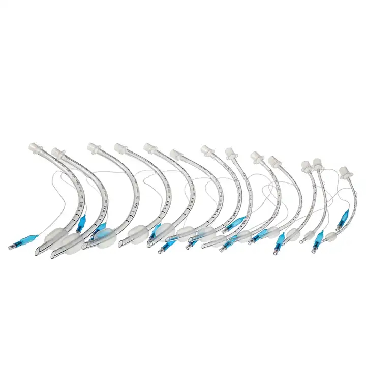
Amoung them, the Standard Endotracheal Tube is a method of inserting a special endotracheal tube into the trachea or bronchus through the mouth or nasal cavity. And the Standard Endotracheal Tube Cuffed is one  type of it, which has different size to adapt to different medical needs,including 3.0mm to 10.0mm.


A cuffed nasal endotracheal tube is a type of nasal tube used for airway management and mechanical ventilation. It is similar to an uncuffed nasal endotracheal tube, but it also includes an inflatable cuff at the distal end of the tube.

Here are some important points about cuffed nasal endotracheal tubes:


  • Design: Cuffed nasal endotracheal tubes are typically made of flexible plastic or silicone material. They have a beveled tip for easier insertion through the nostril and advancement into the trachea. The cuff is located near the distal end of the tube and is inflated once the tube is in the proper position.


  • Cuff inflation: The cuff of a cuffed nasal tube is inflated to create an airtight seal within the trachea. This seal prevents air leakage and reduces the risk of aspiration. The cuff is usually inflated with air using a syringe, and the pressure is monitored to ensure it remains within the recommended range.


  • Advantages: Cuffed nasal endotracheal tubes offer several advantages, including improved ventilation control, reduced risk of aspiration, and the ability to provide positive pressure ventilation. They are commonly used in adult patients, particularly in critical care settings.


Considerations: When using a cuffed nasal endotracheal tube, it's important to carefully monitor the cuff pressure to avoid overinflation or underinflation. Overinflation can lead to tracheal damage, while underinflation may result in air leakage and inadequate ventilation. Regular monitoring and adjustment of cuff pressure are necessary to maintain an appropriate seal.

The specific steps for using a cuffed nasal endotracheal tube are similar to those of other nasal intubation techniques, as described earlier. However, additional attention should be given to proper cuff inflation and monitoring to maintain a secure airway and effective ventilation. Healthcare professionals trained in airway management should follow established protocols and guidelines when using cuffed nasal endotracheal tubes.


Product composition and function:
 
Size10.0mm
Murphy EyeReducing the risk of occlusinon and maintaining airflow
15mm connectorReliable connection to all standard equipment
BalloonProviding even pressure to maintain good sealing,reducing ppressure on the tissues of trachea
Wire coilIncreasing flexibility, providing effective resistance to kinking
ValveEnsuring continual cuff integrity
RadiopaqueAllowing clear identification of the tube on radiographic images
  • Smooth bevelled and carefully moulded hooded tip to assist intubation and to provide high patient safety and comfort.
  • High volume/low pressure cuff helps to ensure an efficient low pressure cuff seal, for intubation during long term ventilation.
  • Intubation depth marks and pre-mounted 15 mm connector.


Endotracheal Tube Cuffed Application Features:
  1. Suitable for both oral and nasal intubation.
  2. Tip-to-Tip X-ray line allows for safe positioning control.
  3. Murphy eye incorporated as an additional safety feature.

Intubation:

  • During intubation, a physician usually stands at the head of the bed looking towards the patient's feet and with the patient lying flat. The positioning will vary depending on the setting and whether the procedure is being done with an adult or child. With children, a jaw thrust is often used.
  • The endotracheal tube with the assistance of a lighted laryngoscope (a Glidescope video laryngoscope is particularly helpful for people who are obese or if a patient is immobilized with a suspected injury to the cervical spine) is inserted through the mouth (or in some cases, the nose) after moving the tongue out of the way.
  • The scope is then carefully threaded down between the vocal cords and into the lower trachea. When it's thought that the endotracheal tube is in the proper location, the doctor will listen to the patient's lungs and upper abdomen to make sure that the endotracheal tube was not inadvertently inserted into the esophagus.
  • Other signs that suggest the tube is in the proper position may include seeing chest movement with ventilation and fogging in the tube. When a doctor is reasonably sure the tube is in position, a balloon cuff is inflated to keep the tube from moving out of place. (In infants, a balloon may not be needed). The tube is then taped to the patient's face.

Verifying Proper Placement:

  1. Once the tube is in place, it's important to verify that it is truly in the proper location to ventilate the patient's lungs. Improper positioning is particularly common in children, especially children who have experienced trauma.
  2. In the field, paramedics have a device that allows them to determine if the tube is in the correct position by a color change.In the hospital setting, a chest X-ray is often done to ensure good placement, though a 2016 review suggests that a chest X-ray alone is inadequate, as is pulse oximetry and physical examination.
  3. In addition to directly visualizing the endotracheal tube pass between the vocal cords with a video laryngoscope, the authors of the study recommended an end-tidal carbon dioxide detector (capnography) in patient's that had good tissue perfusion, with continued monitoring to make sure the tube does not become displaced.
  4. In the setting of a cardiac arrest, they recommended using ultrasound imaging or an esophageal detector device.

After the Procedure:

After the endotracheal tube is in place and a patient connected to a ventilator, healthcare providers will continue to monitor the tubing, settings, and provide breathing treatments and suctioning as needed. Careful attention to oral care will also be provided. Due to the location of the tube, patients who are conscious will be unable to talk while the tube is in place.

Hot Products

PVC Medical Grade murphy eyes with balloon Endotracheal Tube Cuffed Nasal Endotracheal Tube 5.5mm ...
Disposable 6.*7cm Non-Woven Wound Dressing Wound Care Supplier Non-woven adhesive medical dressings ...
10*15CM Surgical Sterile Adhesive Non Woven Wound Dressing with CE,ISO Certificates Non-woven ...
Non-woven Bordered Gauze Island Dressing Wound Care With Absorbent Pads 10*10cm Adhesive Wound ...
Waterproof Non Woven Adhesive Island Dressing Wound Care supplier First Aid Emergency Wound Dressing ...
Transparent waterproof Cohesive Wound Dressings Breathable adhesive wound dressing pad Product ...